

Gastroscopy
Stomach ulcers are now recognised as a major health problem in many horses and can be responsible for a wide range of clinical symptoms, including:
- Weight loss
- Poor appetite
- Decreased performance
- Dull coat
- Mild and recurrent colic
- Stretching to urinate
- Discomfort on girth tightening
- Spending excessive time lying down
- Attitude changes including aggression
- Linked to crib biting.
- No clinical signs at all, while having serious gastric ulcers
Equine Gastric Ulcer Syndrome (EGUS) can affect any horse at any age. Around 90% of racehorses, 60% of performance horses and 35% of non-performance horses are affected, indicating that EGUS is a common problem. Foals and in particular young animals which are being treated for illness or after surgery are at high risk.
A number of factors contribute to EGUS, Exercise and training, diet and in particular long periods without food, stress and medication.
Fortunately, products containing the drug omeprazole which reduces the production of acid in the stomach is curative in 90% of cases in combination with management and feeding practices. Sometimes additional medication is necessary depending on which part of the stomach is involved. Addressing the contributing factors decreases the chances of recurrence of the problem.
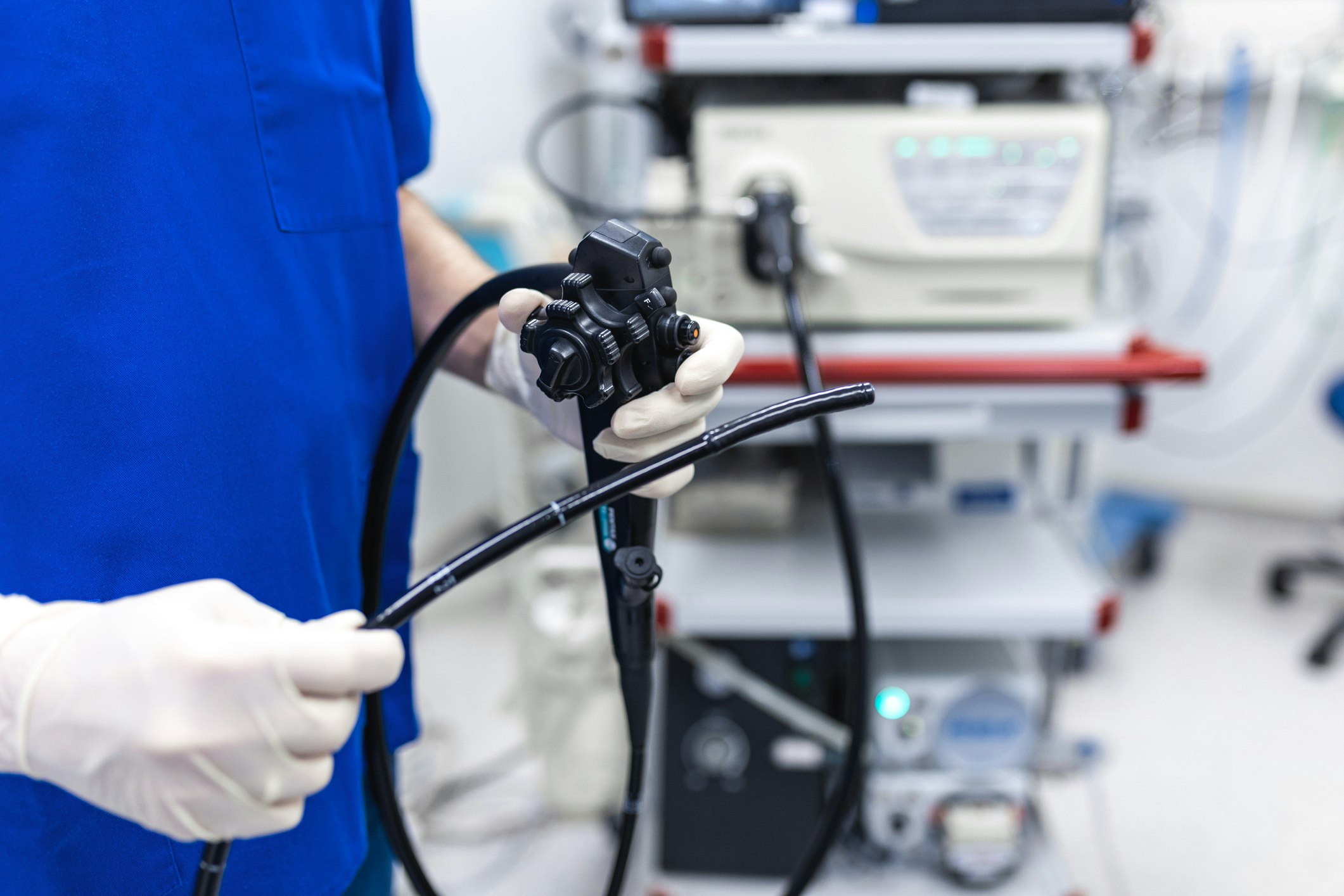
Gastroscopy is the only way to accurately and reliably diagnosing stomach (gastric) ulcers.
Gastroscopy involves passing a 3-meter-long flexible fibre optic camera (gastroscope) up one of the horses nostrils, via the oesophagus into the stomach. Once inside the stomach, the stomach is inflated with air to allow visualisation and full examination of the different regions of the stomach, including the proximal duodenum.
Before scoping a horse must be starved for 12-16 hours. Young foals should not be starved for more than 6 hours. Water is normally withheld up to two hours before scoping.
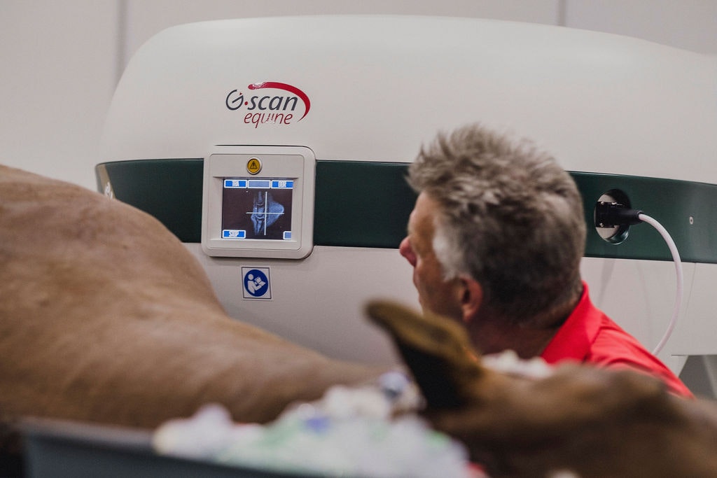
G-scan MRI
The Esaote G-scan is the ultimate MRI system for horses.
The G-scan is the only MRI system capable of scanning the limbs of horses including the carpus, tarsus and stifle. Its design and rotating mechanism allowing to create exceptional three-dimensional imaging, including the head and neck. This technology is the only imaging modality to show bone and soft tissue. MRI can complement lameness evaluations including digital radiography and ultrasonography, diagnosing bone and soft tissue disease and identify very subtle lesions such as bone inflammation.
We welcome referrals from veterinary clinics and their horse clients. Our team strives to be part of the solution and embraces a team approach to providing a definitive diagnosis.
Directions
Drive straight through the main gate, stay on this road for approximately 1km. At the intersection at the top of the hill turn right at the 'Tree of Paradise' sculpture (red tree sculpture) and follow the road past the arenas and down the hill to the Vet Hospital.
Contact
Willinga Park Vet Hospital
02 4405 5658
vethospital@willinga.com.au
Share this page
Newsletter Signup
Add your email address & subscribe to receive our newsletter